Asto CT Honors 2024's Best CT Cases: Showcasing Outstanding Equine Diagnostics
We are thrilled to announce the results of this year’s Asto CT Best Scans of the Year Contest! A heartfelt thank you to all participants for submitting such outstanding and diagnostically valuable cases. This year's entries highlight exceptional advancements in 3D imaging, head scans, limb scans, and soft tissue filter scans. Below are the final standings by category, featuring our first-place winners and overall highpoint winner.
Limbs Category
Vet CT Judging Comments: Higher marks were awarded to cases where CT studies offered clear diagnostic value beyond traditional imaging techniques.
Featured Winner
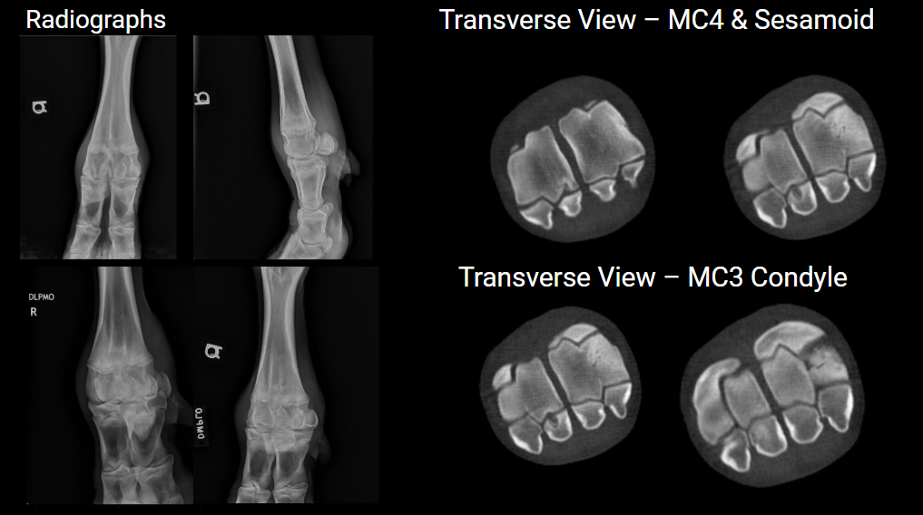
First Place: Dr. Brenda Righter, University of Wisconsin – Osteochondritis Dissecans
Highest-scoring entry with a score of 31
Recognized for outstanding case selection, exceptional image quality, comprehensive documentation, and significant clinical impact, including lesion detection, thorough assessment, and pre-surgical planning.
History: 11mo Holstein bull (390kg) presented for 1 week history of lameness and fetlock swelling
Presentation:
4/5 lame on the right front limb
Marked RF fetlock effusion
Mildly tender to palpation, no heat noted
Remainder of exam WNL
Diagnostics:
Ultrasound – Moderate anechoic intracapsular effusion, osseous roughening of the medial aspect of metacarpal 3 condyle noted
Arthrocentesis
Synovial Cytology
Microbiology (Synovial fluid)
Aerobic Culture
Anaerobic Culture
Mycoplasma Culture
Mycoplasma spp PCR
All negative after 7 days
Advanced Imaging
Radiographic Interpretation
Soft tissue swelling of distal metacarpus, most severe plantar aspect
Normal cutback zone distal 3rd/4th metacarpus
Mild joint effusion/synovial hyperplasia
Findings deemed inconsistent with severity of lameness, particularly with low indication of infectious process. Elected to proceed with standing CT evaluation.
Diagnosis
Osteochondrosis Dessicans
Cystic lesion of medial proximal sesamoid bone of digit 4 with corresponding lytic lesion of MC4
Multiple subchondral defects of the condyles of MC3
Patient underwent arthroscopic debridement of observed osteochondral lesions.
Outcome
Patient recovered uneventfully from surgery.
Post-op care managed in hospital. Suture removal 14d post-op.
Patient doing well at home and owners have been successfully collecting him.
Head and Neck Category
Vet CT Judging Comments: Higher scores were given to cases where CT studies demonstrated diagnostic value beyond traditional imaging.
Featured Winner
First Place: Dr. Douglas Langer, Sara Clark, Dr. Lindsay Monroe, Dr. Kari Means, and Dr. Jennifer Gold, Wisconsin Equine Clinic and Hospital – Severe Degenerative Injury with Joint Space Collapse at C1-C2
Recognized for exceptional image quality, innovative case selection, and critical diagnostic insights contributing significantly to case management and clinical decision-making.
Signalment:
10-year-old Arabian Saddlebred cross mare
History:
Patient was referred for evaluation of the head and neck after the referring veterinarian identified an abnormal noise and palpable movement in the cranial cervical region. On examination, the mare did not exhibit signs of pain, though a loud, dull noise was noted during neck movement, originating in the upper cervical area. The mare did not display any protective posturing or abnormal head positioning.
Diagnosis:
Severe degenerative injury and joint space collapse at the C1-C2 articulation, accompanied by extensive subchondral and trabecular bone abnormalities, indicating permanent structural damage to the joint.
3D Category
Updated First Place Winner
Congratulations to Wisconsin Equine Clinic and Hospital for their case, Severe degenerative injury with joint space collapse at C1-C2, earning a score of 24. See above for case information.
Vet CT Judging Comments: Higher marks were reserved for cases where the 3D reconstructions demonstrated clear diagnostic value beyond the 2D dataset. These cases remain rare in equine imaging.
Featured winner
First Place: Dr. Sabrina Brounts, University of Wisconsin-Madison — 3D Sequestrum
Awarded first place due to excellent anatomical coverage, good image quality, lack of imaging artifacts, and additional procedure involving the use of contrast imaging.
History: 13-year-old Shire gelding
Presented with a 10-month history of right front limb lameness.
Physical Exam:
Lameness: IV/V on AAEP scale, right front limb.
Thickening on the lateral aspect of the hoof near the coronary band.
Draining tract with mucopurulent discharge on the lateral aspect of the hoof near the coronary band.
Imaging: CT scan with contrast performed.
Findings: Sequestrum and involucrum in the right lateral palmar process of the distal phalanx with surrounding osteitis.
Draining Tract: Leads to sequestrum.
Treatment: Standing surgery for removal of sequestrum and excision of draining tract.
Outcome: Horse recovered well from surgery.
Soft tissue category
Vet CT Judging Comments: Higher marks were awarded to cases where soft tissue filter images added diagnostic value beyond bone filter and ancillary imaging.
Featured Winner
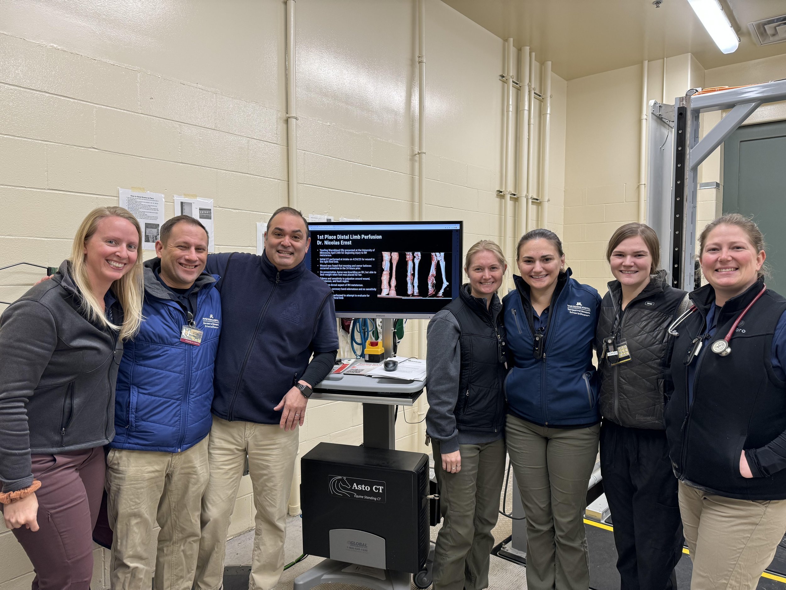
First Place: Dr. Nicolas Ernst, Shannon Vesledahl, University of Minnesota — Bilateral Navicular Disease
Awarded first place due to clear visualization of DDFT soft tissue lesion and diagnostic value in the soft tissue and bone filter images. Entry also provided significant documentation and information.
Patient History/PE:
5-year-old QH mare with bilateral forelimb lameness.
Previous diagnoses: navicular syndrome and left front navicular bone cyst.
Coffin joint injections (steroid and arthramid) no longer effective.
Responds to anti-inflammatories but not currently on any.
Exam findings:
3/5 LF lame.
Positive to distal limb flexions of bilateral forelimbs.
PD nerve block on LF switched lameness to 1/5 RF.
Abaxial block on LF resulted in 2-3/5 RF lameness.
Diagnosis:
Bilateral podotrochlear apparatus abnormalities:
Moderate to marked navicular bone changes (greater on LF).
LF deep digital flexor tendinopathy with dorsal border tears near navicular bursa.
Mild bilateral deep digital flexor and impar enthesopathy.
Mild bilateral distal interphalangeal joint effusion.
Subtle subchondral contour defect in RF distal P2.
Minimal bilateral fetlock osteoarthritis.
Treatment/Outcome: Owner elected to discuss retirement options for the mare.
Highest Scoring Clinics
Congratulations to our highest scoring clinic University of Minnesota-Leatherdale Equine Clinic.
Contest Participants
Dr. Nicolas Ernst, DVM, MS, Diplomate ACVS – University of Minnesota
Shannon Vesledahl, BS, CVT – University of Minnesota
Dr. Kevin G. Keegan, DVM, MS, Diplomate ACVS – University of Missouri
Dr. Sabrina H. Brounts, DVM, MS, PhD, Diplomate ACVS, ECVS, ACVSMR – University of Wisconsin-Madison
Dr. Brenda Righter, DVM – University of Wisconsin-Madison
Dr. Douglas Langer, DVM, MS – Wisconsin Equine Clinic and Hospital
Sara Clark, CVT – Wisconsin Equine Clinic and Hospital
Dr. Lindsay Monroe, DVM – Wisconsin Equine Clinic and Hospital
Dr. Kari Means, DVM – Wisconsin Equine Clinic and Hospital
Dr. Jennifer Gold, DVM – Wisconsin Equine Clinic and Hospital
Dr. James Whitmore, BVetMed CertAVP MRCVS – Cotts Equine Hospital
Rahim Ahammed – Sharjah Equine Hospital
Dr. Naroa Arteagoitia, DVM – Sharjah Equine Hospital
Judging Criteria
Clarity and Resolution: High-resolution images free of artifacts and with clear, detailed anatomy.
Diagnostic Value: The scans’ ability to reveal significant findings.
Innovative Cases: Unique or challenging cases highlighting the versatility of CT.
Clinical Impact: Influence on treatment, prognosis, or case management.
Anatomical Coverage: Thorough evaluation of relevant anatomical structures.
Positioning and Technique: Optimal positioning and correct CT technique.
Documentation and Information: Completeness of case details provided with the scans.
Congratulations to all winners and participants for showcasing exceptional clinical cases and advancing the role of CT imaging in equine diagnostics. The Asto CT team expresses our gratitude to VET CT for their expertise in judging all 50 entries. Each deserving winner received a $100 Amazon gift card, Stanley Cup, and exclusive Asto CT and VET CT merchandise. We look forward to seeing even more innovative and impactful submissions next year!


