We're pleased to announce significant progress in deploying our technology to image the cervical spine. We were able to reach the cranial aspect of C7 at Virginia Equine Imaging on several standing patients.
Read More
We're pleased to announce significant progress in deploying our technology to image the cervical spine. We were able to reach the cranial aspect of C7 at Virginia Equine Imaging on several standing patients.
Read More3 yr old Gypsy Vanner gelding was referred for evaluation of a persistent left facial swelling that had been present for five months.
Read More5 yr old TB gelding presented following an acute episode of severe pain, during which he went down and thrashed.
Read More15 yr old Saddlebred with a 1- to 2-month history of right forelimb (RF) lameness.
Read More8 yr old warmblood gelding, concern for lateral collateral ligament involvement or possible OCD in the left hock.
Read MoreA 13-year-old Gypsy mare was brought in with a several-month history of a protuberance from the right maxilla, which had recently started to drain. Radiographs indicated…
Read More5-year-old Quarter Horse mare History: Presented with bilateral forelimb lameness; previous diagnoses include navicular syndrome and left front navicular bone cyst.
Read MorePatient was referred for evaluation of the head and neck after the referring veterinarian identified an abnormal noise and palpable movement in the cranial cervical region.
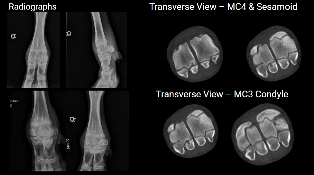
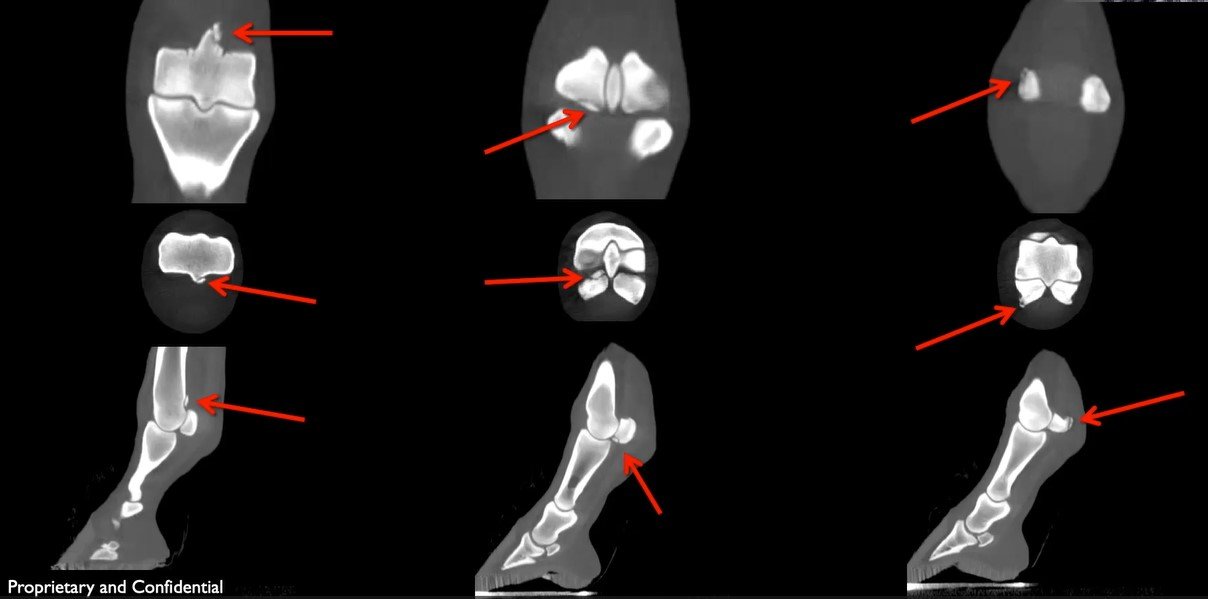
Read MoreThe CT identified osteochondritis dissecans, including a cystic lesion in the medial proximal sesamoid bone, lytic lesions in MC4, and subchondral defects in MC3.
Read More5-year-old POA mare presented at the University of Minnesota for CT of the right front foot. She has an approximately 1-year duration of Grade 2/5 right front lameness.
Read More9-month-old Percheron gelding presented for respiratory noise, leading to the discovery of a multilocular aneurysmal bone cyst.
Read MoreA yearling Warmblood filly presented for a degloving injury to RH metatarsus
Read More16 year old Argentinian Warmblood Gelding presenting at the University of Minnesota for 4/5 right front lameness for a 10-14 days duration after 2 incidences of trauma (fighting with other horses).
Read More24yo Arabian cross mare presented for unilateral left nasal discharge unresponsive to antibiotics.
Read More24 yr old gelding, with non-healing puncture wound to LF foot.
Read More4yo QH stallion, Acute non-weight bearing lameness following a gallop.
Read More10 year old Tennessee Walking Gelding presented to the University of Minnesota for facial swelling.
Read More5 yr old Quarter Horse with a front limb lameness associated with a 3 cm laceration.
Read MorePercheron draft gelding with chronic right front limb lameness going on for at least a year.
Read MoreIn this video the Equina imaged a tendon lesion, squamous cell carcinoma, and neurologic disorder in the neck.
Read More