Osteochondritis Dissecans


Featured Winner: Dr. Brenda Righter, University of Wisconsin – Osteochondritis Dissecans
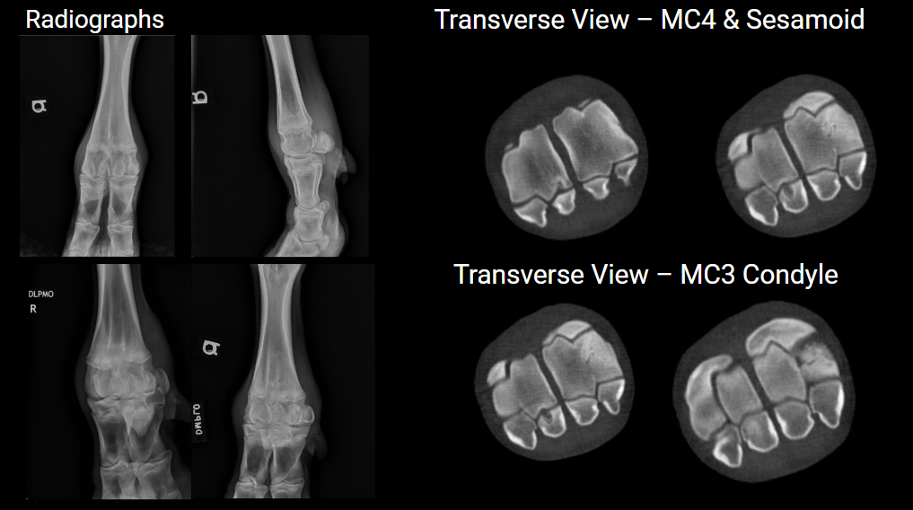
Dr. Brenda Righter earned first place with the highest-scoring entry (31 points) for her outstanding case featuring an 11-month-old Holstein bull (390kg) with a one-week history of lameness and fetlock swelling. Recognized for exceptional image quality, thorough documentation, and significant clinical impact, her work highlighted the case’s diagnostic and surgical journey.
Case Summary:
The bull presented 4/5 lame on the right front limb, with marked fetlock effusion and mild tenderness but no heat. Initial diagnostics, including ultrasound and synovial fluid analyses, suggested moderate effusion and medial condyle roughening but no infection.
Advanced Imaging:
Radiographs revealed inconsistencies with the lameness severity, prompting a standing CT scan. The CT identified osteochondritis dissecans, including a cystic lesion in the medial proximal sesamoid bone, lytic lesions in MC4, and subchondral defects in MC3.
Treatment and Outcome:
Arthroscopic debridement addressed the lesions, and the bull recovered uneventfully with post-op care managed in-hospital. After suture removal at 14 days, the patient returned home in good health, with the owners successfully collecting him.
