THE EQUINA® BY ASTO CT
BUILT FROM THE GROUND UP, THE EQUINA IS THE WORLD’S ONLY TRUE WEIGHT-BEARING, FAN-BEAM CT SCANNER WHICH ALLOWS FOR SCANNING IN A NATURAL STANDING POSITION.

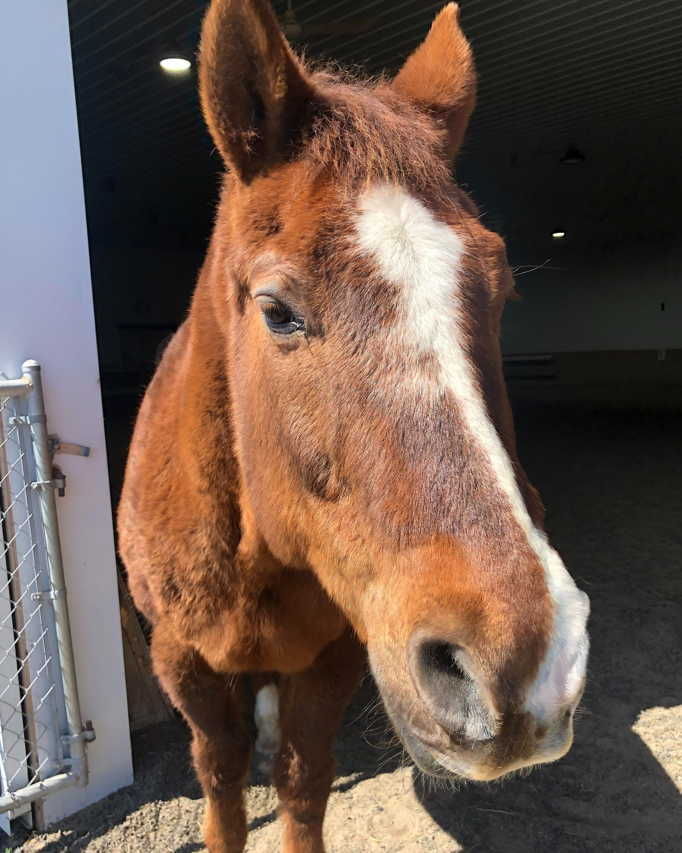
HI, MY NAME IS CHESTER
Chester is a retired show horse that currently serves as a decorative pasture companion. His owner made the decision to bring him to the clinic due to emerging complications within his nasal cavity.
CLINICAL EXAMINATION
Chester presented to the clinic with nasal discharge and intermittent epistaxis (nosebleed). He was subsequently recommended for a head CT scan in order to further investigate the underlying cause of his symptoms.
CT FINDINGS - ETHMOID HEMATOMA
CT images show a soft tissue mass in the left nasal cavity. This mass is intermittently associated with the left ethmoid turbinate and surrounded by soft tissue that has fluid attenuation and fills the surrounding sinuses. Right side is normal.
WHAT IS AN ETHMOID HEMATOMA?
An ethmoid hematoma is a well-recognized but poorly understood disease in horses. Affected horses classically exhibit bloody nasal discharge originating from a hematoma-like mass within the ethmoid turbinates of the nasal cavity. source
CT VIDEO: In the region of the mass a hyperdense swirling appearance can be seen which is highly suggestive of an ethmoid hematoma.
SURGERY
The mass was removed through a frontonasal flap. The 3D reconstruction shows staples and sutures that closed the flap. Chester is reportedly doing well at home. Histopathology – findings consistent with ethmoid hematoma with bacterial colonies.
WHY IS CT USEFUL?
CT is useful in ethmoid hematoma cases where the mass cannot be viewed endoscopically because of the size or radiographically. CT scans provide a three-dimensional view of the hematoma and its relationship with adjacent structures. This helps surgeons plan the most appropriate treatment approach, whether it involves surgical removal or other therapeutic options.
Find Out More About The Equina® CT Scanner
CLICK THE ICONS BELOW
Find a CT location near you
Other featured case studies
A WORD FROM OUR SUPPORTIVE EQUINE VETERINARIANS

Stay in the loop with Asto CT


