

THE EQUINA® BY ASTO CT
BUILT FROM THE GROUND UP, THE EQUINA IS THE WORLD’S ONLY TRUE WEIGHT-BEARING, FAN-BEAM CT SCANNER WHICH ALLOWS FOR SCANNING IN A NATURAL STANDING POSITION.


HI, MY NAME IS MABEL
Mabel is a 5-year-old Quarter Horse mare who runs barrels. Her owner made the decision to bring her to the clinic due to a traumatic injury that occurred in her stall overnight. She had a severe lameness in her left hind leg, and a wound near her fetlock area.

CLINICAL EXAMINATION
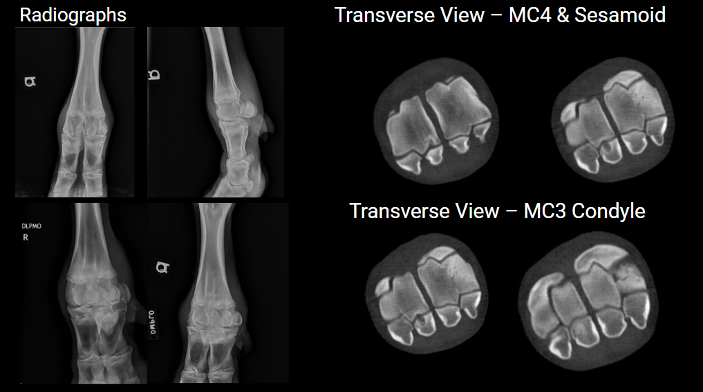
Upon initial examination, clinicians took radiographs which showed the presence of several fracture fragments in her fetlock area and swollen soft tissue of the distal limb. Subsequently she was recommended for a CT scan to further investigate in more detail.
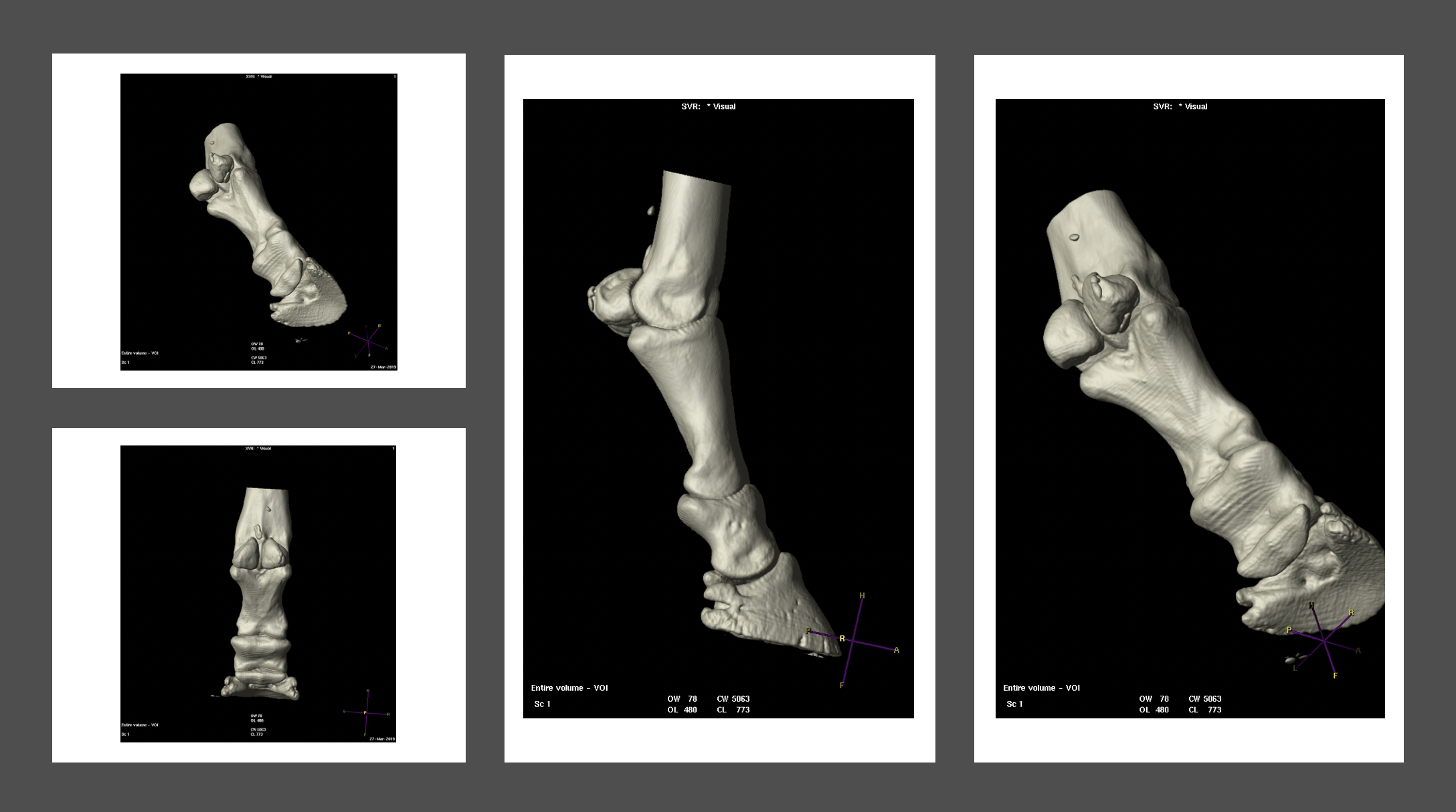
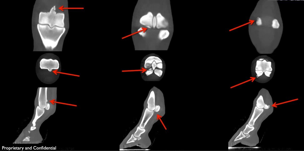
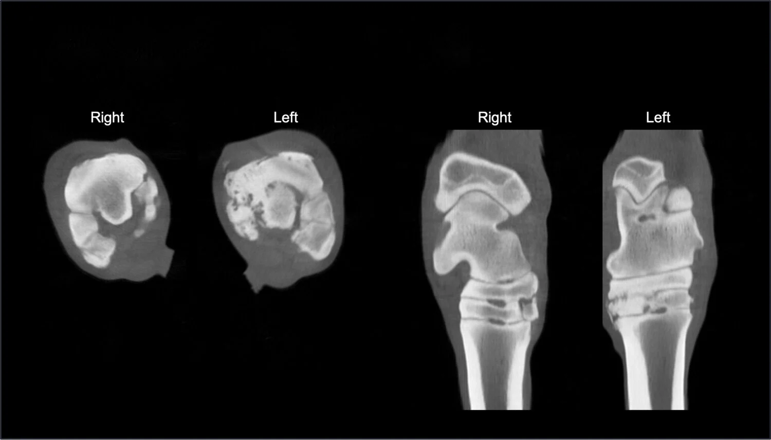
CT FINDINGS - MULTIPLE FRACTURES FRAGMENTS
CT images showed acute, traumatic, complete, comminuted fractures involving the medial proximal sesamoid bone (basilar and abaxial) and third metatarsal bone (sagittal ridge). Additionally, an intracapsular metatarsophalangeal fracture fragment with effusion of the fetlock joint was noted. This condition was challenging to diagnose due to the presence of multiple fractures in the fetlock region.

PROXIMAL SESAMOID FRACTURES
Proximal sesamoid fractures range from simple to complex, and may be articular as well as non-articular. These types of fractures are commonly seen in racehorses due to hyperextension type injuries; however, in this case trauma was the cause of the injury. When a proximal sesamoid fracture occurs, it can result in lameness and discomfort for the horse. They can potentially disrupt the function of the surrounding tendons and ligaments, which are crucial for the horse's movement and support.

SURGERY
Mabel was treated surgically with arthroscopy to remove the fragments and debride the traumatic wound.
WHY IS CT USEFUL?
Asto CT's technology allows veterinarians to obtain three-dimensional images of the horse's limb while the animal remains standing, eliminating the need for anesthesia and providing a more comprehensive view of the injury. This precise imaging was instrumental in diagnosing Mabel’s acute fractures, determining the extent of the damage, and identifying the best treatment plan with minimal complications. With this information, veterinarians can develop surgical and treatment plans individualized to their patient and improve the chances of a successful recovery.
CT Video: Shows multiple fracture fragments in a 3D reconstruction and MPR scan.
What Areas Can the Equina® scan?
Head and Upper Neck To C5 standing
Caudal Neck Under General Anestesia
Stifles Under General Anestesia
Front and hind limb pairs-
knee/hock to the foot standing
Find Out More About The Equina® CT Scanner
CLICK THE ICONS BELOW


Other featured case studies


A WORD FROM OUR SUPPORTIVE EQUINE VETERINARIANS

Stay in the loop with Asto CT
